
Finished! Looks like this project is out of data at the moment!
We've just added a brand new workflow to Etch A Cell - ImmunoExplorers! Check out 'Workflow 2 - Nuclei Hunters'! Thanks for being here 😃
Research
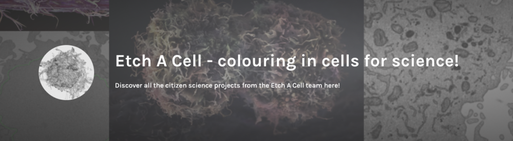
Welcome to Etch A Cell - ImmunoExplorers
Who are we, what are we studying, and why?
The science team behind Etch A Cell are based at the Francis Crick Institute in London, UK. There they work with many research teams to study different aspects of biology using cutting-edge Electron Microscopes. These microscopes have a very high magnification and resolution, and so can be used to take highly-detailed images of many things, including cells, molecules and tissues. These images can be used to provide us with a richer understanding of biology, which can help us understand the biological changes associated with health and disease.
Recent developments in electron microscopy mean it is possible to collect images automatically, so our team is now producing a huge amount data; looking at a single cell alone can produce several terabytes worth! Although these technological advances mean we can perform our research much faster, this flood of data has caused a data analysis bottleneck, which is why we need your help!
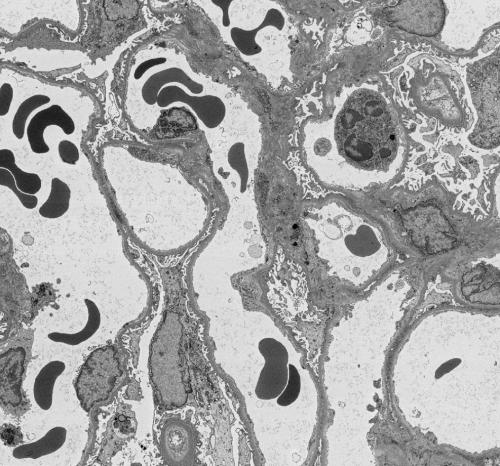
In this project you'll get to see some very high resolution images of kidney tissue, like this one!
How you can help – colour in cells for science!
To make sense of the images taken by the electron microscope we must carefully analyse them. To extract meaning from the images we segment the features we’re interested in, which means to draw around the bits of the image that we want to study. We need your help with this task – to colour in different parts of the image. In this Etch A Cell project, we would like you to help us to study immune cells in kidney tissue.
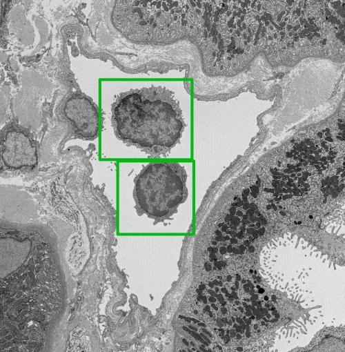
In this project we need you to spot and draw around immune cells, which you can see in this image outlined in green (for more examples, see our immune cell gallery in the Field Guide tab which you can find on the right hand side of your screen)
Why are we studying immune cells in kidney tissue?
Chronic kidney failure is a significant health issue that impacts about 11% of people worldwide, with diseases caused by the immune system being the third most common cause of chronic kidney failure. When someone's kidneys reach the point of failing, the most effective treatment currently available is a kidney transplant. Unfortunately, the lifespan of a transplanted kidney typically averages only 10 to 15 years. This is mainly due to the recipient's immune system responding negatively to the donated kidney, a condition known as ‘transplant rejection’.
Our current understanding of rejection comes from examining biopsies of the transplant using a standard light microscope, and from analysing expression of genes in biopsy tissue. Antibodies and other molecules gather in the small blood vessels of the kidney and attract a variety of immune cells. These immune cells get 'activated' and damage the lining of the blood vessels, causing thickening of the vessel walls. Eventually, this prevents the kidney from filtering fluid and waste from the blood.
What are we hoping to learn from this project?
There is still a lot we don't know about kidney transplant rejection; we have a lot to learn about which immune cells are involved, how they get activated, and what effect this has on the lining of the blood vessels of the kidney. This is partly because it is difficult to study the molecules and cells within the small blood vessels in human biopsies taken for diagnosis. One of the biggest challenges in studying tissues at high resolution is how long it takes – to image a standard-sized biopsy would take up to 55 years for a single sample!
Through our research, we’re hoping to reduce the time taken to analyse a human biopsy from 55 years to 5 days. At this speed, we will be able to amass data from enough biopsies that we can start to look for patterns in the recruitment of antibodies and immune cells to the kidneys of patients with transplant rejection. Ultimately, we will be able to use this information to predict which transplants will fail and to inform the development of new treatments of organ rejection.
Through contributing to this Zooniverse project, you’re helping to speed up our data analysis pipelines!
Why do we need your help?
The process of manually segmenting (drawing around) these structures can take a really long time - it is the major bottleneck in our research - this is why we need your help! While it may look as though computers would be able to perform this process very well already, in practice they are still quite error-prone and the time it takes to perfect the algorithm can be longer than the time to do the segmentations by hand. Small variations in the images, either due to real biological differences or due to slightly different imaging conditions, often render a finely-tuned algorithm ineffective for other data, sometimes meaning that the algorithms need to be optimised for each and every sample!
We hope that this project will advance our ability to analyse biological features using electron microscopy and segmentation. With enough volunteer help we may be able to train computers to segment automatically; this has huge potential to help us understand biology and study disease!
Further information
For more information about the scientific methods we use in our research, take a look at the FAQ.
This project is part of the Etch A Cell Organisation
'Etch A Cell - ImmunoExplorers' is one of multiple projects produced by the Etch A Cell team and their collaborators to explore different aspects of cell biology. If you'd like to get involved in some of our other projects, you can find the other Etch A Cell projects on our organisation page here.
With thanks to support from...


Thanks for contributing to Etch A Cell - ImmunoExplorers!