


Please give us your feedback using this short Google form https://forms.gle/EpUNtjv76qMtReSo6
Research
Overview of our research
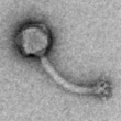
We are researchers at the School of Life Sciences at the University of Warwick, where we help other researches to use microscopes to investigate their samples. These can be anything from bacteria to brains. Some of the researchers here work on antibiotic resistant bacteria and bacteriophage therapy. Bacteriophage are viruses that infect and kill bacteria and people have been excited about using these as treatment for infections, especially as antibiotics are getting less effective. People look for bacteriophage in many places, such as polar ice caps, deep-sea hydrothermal vents and volcanic hot springs. But we're looking in ponds.
Viruses are everywhere!
This project started with a visit from schoolchildren in their summer holidays, where they looked at the diversity of microscopically tiny things in the pond outside our building, both with light microscopes, and with the electron microscope. We were inspired by the weird and wonderful things we found.
Few people realise the mind-blowing abundance of viruses in our environment: they outnumber every living thing on the planet. Every day around 800 million land on every square meter of the earth, a millilitre of seawater can contain tens of millions of them, the local pond is basically viral soup. To find out more, read the comic
We want to raise public awareness of viruses – specifically bacteriophage, just how common they are and how important they are both as potential disease treatments and as vectors for spreading antibiotic resistance and disease causing genes.
This project is special because members of the public can provide samples AND help with data analysis. The results will be used to build up a database of morphology by post code and water type to look for any correlations. We also ask volunteers to just keep an eye out for anything unusual: we’ve found phage normally associated with hot springs and several species of giant virus. We are continuously looking for more funding to be able to process more samples and get some genetic information on the most interesting ones.
Our scientific methods
When we receive the water sample, we use a paper filter to get all the big things out. We then collect all the things we're interested in by spinning the sample in an ultracentrifuge. This sends everything in the water - viruses, small bacteria, tiny pieces of clay and sand - to the bottom of the tube. We mix it in with a tiny bit of water again and apply it to a grid. We then use a heavy metal stain to make the viruses and other things visible in the electron microscope. You can read more about our sample preparation methods here.

Why do we need your help?
We are getting many samples in from schools in Coventry and the surrounding area. For each sample, we can take many many images and each of these images may have something exciting in it! Because of the many different shapes in the images, computers are not very good at telling us where the particles are, and that's where you come in. The human brain has evolved to see patterns in everything, and this is exactly what we need.
What are our research goals?
- See if certain virus types (shapes) are more likely to show up in certain types of water, such as rivers, ponds or canals.
- See if some previously undescribed shapes from our pond occur in other water sources
- See how common giant viruses are in the UK