


Finished! Looks like this project is out of data at the moment!
Congratulations on completing another dataset! We're so appreciative of your incredible work!
Research
Overview of Etch A Cell - ER
Who are we, what are we studying, and why?
The science team behind Etch A Cell are based at the Francis Crick Institute in London, UK. There they work with many other researchers to study different aspects of biology using cutting-edge Electron Microscopes. These microscopes have a very high magnification and resolution, and so can be used to take highly-detailed images of many things, including cells, molecules and tissues. These images can be used to provide us with a richer understanding of biology, which can help us understand the biological changes associated with health and disease.
Recent developments in electron microscopy mean it is possible to collect images automatically, so our team is now producing a huge amount data; looking at a single cell alone can produce several terabytes worth! Although these technological advances mean we can perform our research much faster, this flood of data has caused a data analysis bottleneck, which is why we need your help!

In this project you'll get to see some very high resolution images of cells like this one!
How you can help – colour in cells for science!
To make sense of the images taken by the electron microscope we must carefully analyse them. To extract meaning from the images we segment the features we’re interested in, which means to draw around the bits of the cell that we want to study. We need your help with this task – to colour in different parts of the cell. In this Etch A Cell project, the cellular feature we would like you to help us segment is the endoplasmic reticulum (ER).
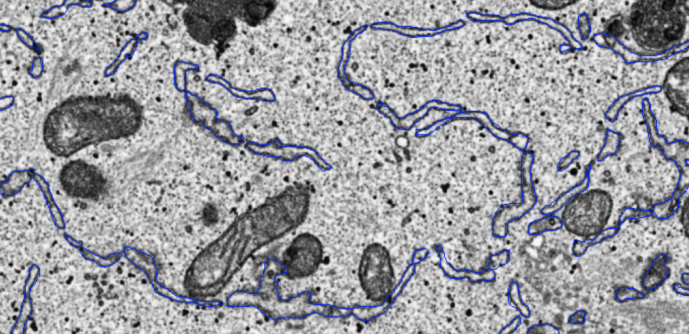
In this project we need you to spot and draw around the endoplasmic reticulum, which you can see in this image outlined in blue (for more examples, see our ER gallery in the Field Guide tab which you can find on the right hand side of your screen)
The Endoplasmic Reticulum – one of the biggest structures in your cells!
The endoplasmic reticulum is a type of organelle (a specialized structure) found within most eukaryotic cells. It is often one of the biggest organelles in the cell, and is made up of an interconnected network of membranes (called 'cisternae') that can take a variety of shapes and sizes. The endoplasmic reticulum plays a role in many important cellular functions including the production of essential cell building blocks such as fats and proteins. In this project, we ask you to draw around any endoplasmic reticulum you can spot in each image. This effort will help significantly improve our understanding of endoplasmic reticulum biology.

The endoplasmic reticulum is a large organelle found in most eukaryotic cells. It's formed from a network of membranes ('reticulum' means 'network'!)
Why do we need your help?
The process of manual segmentation can take a really long time - it is the major bottleneck in our research - this is why we need your help! While it may look as though computers would be able to perform this process very well already, in practice they are still quite error-prone and the time it takes to perfect the algorithm can be longer than the time to do the segmentations by hand. Small variations in the images, either due to real biological differences or due to slightly different imaging conditions, often render a finely-tuned algorithm ineffective for other data, sometimes meaning that the algorithms need to be optimised for each and every sample!
We hope that this project will advance our ability to analyse biological features using electron microscopy and segmentation. With enough volunteer help we may be able to train computers to segment automatically; this has huge potential to help us understand biology and study disease!
Further information
For more information about the scientific methods we use in our research, take a look at the FAQ.
In this project we're working with friends!
Some of the data sets you will work on in Etch A Cell - ER are of interest to other researchers beyond the core Etch A Cell team. You can read a little bit more about their projects below:
Jez and Guy - studying how the endoplasmic reticulum changes when cells divide
Cell division is essential for life, growth and development. When cell division goes wrong, it can lead to the development of diseases such as cancer and developmental abnormalities. While we know that DNA is contained within a cell’s nucleus, cells are also stuffed full of intracellular compartments called organelles. DNA division is well understood, but we know far less about how these organelles are split equally between the daughter cells, and how they regain their function after division. We are particularly interested in a massive organelle called the Endoplasmic Reticulum that changes its shape during division. We’ve used the Francis Crick’s state-of-the-art microscopes to take some ultra-high resolution images of all the organelles inside dividing cells at difference stages of cell division. This microscopy paints amazing, but incredibly complex, 3-dimensional pictures. We need your help to extract the relevant organelles from the data. This will allow us to build a cutting-edge 3-dimensional picture of how these organelles change shape as cells divide.

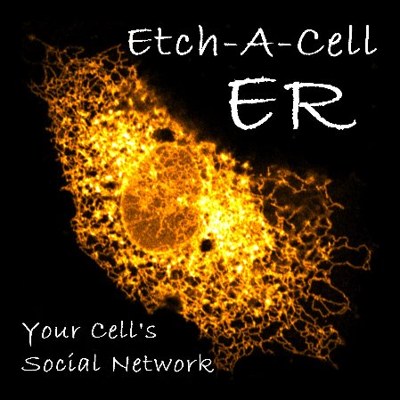
A beautiful image of the endoplasmic reticulum from Jez and Guy's research
This project is part of the Etch A Cell Organisation
'Etch A Cell - ER' is one of multiple projects produced by the Etch A Cell team and their collaborators to explore different aspects of cell biology. If you'd like to get involved in some of our other projects, you can find the other Etch A Cell projects on our organisation page here.
With thanks to support from...


