


Thank you everyone for making this project a great success! You have helped us successfully complete our current data set! We are planning to upload new data here soon! Please keep any eye out for our announcements on Talk!
This experience works best on desktops and laptops which have mouse or track-pad capabilities. We recommend using one of these for the best results!
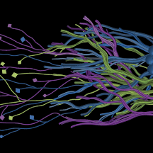
Warning: This project shows 3-dimensional data of brain tissue from non-human primates imaged using fluorescence microscopy.
Research
Mind Mapper: Mapping the Brain’s Hidden Connections
Unraveling the Brain’s Circuitry
Understanding how different parts of the brain connect is one of the biggest challenges in neuroscience. Scientists have found that specific circuit abnormalities are linked to various brain disorders, making it important to map these connections in detail. This project focuses on creating a high-resolution connectivity map, or "Connectome," between the frontal and parietal lobes—two key regions of the mammalian brain involved in thinking and decision-making.

How Do Scientists Map Brain Connections?
The basic unit of a brain circuit is the axon - the pathways that carry information and connect different neurons within the brain. Researchers use several imaging techniques to study these axons, or brain circuits, each capturing different levels of detail:
- Diffusion Magnetic Resonance Imaging (dMRI): Provides a large-scale view of axon bundles (hundreds to thousands of micrometers) by analyzing how water moves through brain tissue.
- Polarization-Sensitive Optical Coherence Tomography (PS-OCT): Captures smaller axonal structures (a few micrometers) by detecting light polarization changes in the myelin sheath (the protective covering around axons).
- Tract Tracing: Uses glowing viral particles to trace individual axons, offering the highest resolution but requiring invasive procedures and long processing times.
Each method has its strengths and challenges. While dMRI is fast, it has low resolution. Tract tracing provides incredible detail but takes years to complete and involves complex procedures.

(Check the FAQ page for more details and definitions for terms commonly used in neuroscience!)
A Faster, Less Invasive Approach
To overcome these challenges, scientists are exploring ways to infer small-scale axonal connections using non-invasive imaging like dMRI. Imagine predicting fine details about individual axon pathways using a “blurrier” large-scale image! If successful, this approach could revolutionize neuroscience by accelerating research and improving our understanding of brain disorders.
Your Role in this Groundbreaking Research
At the Center for Mesoscale Connectomics (CMC), researchers have developed a new strategy that combines all three imaging techniques—dMRI, PS-OCT, and Tract Tracing—using data from non-human primates, whose brain structures closely resemble those of humans. By correlating different imaging results, they aim to create a comprehensive axonal connectome for both primates and humans.
However, analyzing this data is a massive challenge. While machine learning can assist, human expertise is still needed to verify the results. That’s where you come in!
Join the "Mind Mapper" Project!
We are launching Mind Mapper on Zooniverse, where you can help annotate axonal tracts within 3D brain imaging data. You’ll use our new Volumetric Subject Viewer to explore and interact with real brain structures in 3D. To make things even easier, we’re introducing a semi-automated annotation tool to help you work faster!
Your contributions will directly support cutting-edge neuroscience research, bringing us closer to understanding brain connectivity and its role in neurological disorders.
We are excited to have you on this journey—thank you for being part of this groundbreaking project! 🚀🧠
A transparent disclosure to our volunteers: The Mind Mapper project follows all federal and institutional guidelines for the ethical care and use of animals in research. The work is approved by the Institutional Animal Care and Use Committee (IACUC) at the University of Minnesota. This project operates under Animal Welfare Assurance number D16-00288 (A3456-01), which ensures compliance with the policies described by the Public Health Service Policy on Humane Care and Use of Laboratory Animals. Overall our project will use existing brain imaging data collected at the University of Minnesota under the above mentioned approved research protocols. This means no new animal experiments are being performed specifically for this Zooniverse project. For initial calibration data, we are using publicly available products from this site. We are looking to you, our volunteers, to help us turn existing data into new discoveries about the inner workings of our brains🧠!