


Finished! Looks like this project is out of data at the moment!
Thanks to the amazing dedication of our volunteer community, this project is now finished!
You can learn more about Etch A Cell and discover our new projects here
Research
Overview of our research
Our research group works at the Francis Crick Institute in London, where we use a range of cutting-edge Electron Microscopes to take images of molecules, cells and tissues. We collaborate with many scientific researchers to image a huge variety of biological samples to help understand cancer, infectious diseases (including HIV, tuberculosis, malaria), the immune system, the brain and nervous system, diabetes and more. We collect images of these samples using our electron microscopes, some of which are automated. This means we can produce a vast amount of image data; one cell alone may produce up to several terabytes worth of data!
After producing this data, the next step is to analyse and understand it - which is where we need your help! To extract meaning from the image data we produce, we need to “segment” the images; this means to draw around the cell features of interest.
In this project, the feature of interest is the nuclear envelope. The nuclear envelope is the barrier that seperates the genetic information (DNA) inside the nucleus from the chemicals and reactions going on in the rest of the cell. The nuclear envelope is a fascinating structure. It is able to break down every time the cell divides, and then reforms again to protect the DNA in the new daughter cells. It also acts as a gatekeeper, tightly regulating the molecules that are allowed to pass in and out of the nucleus. However, because it has such an important role in the cell, problems with the nuclear envelope can cause a range of different diseases. Changes in the shape of the nuclear envelope may be involved in common diseases like virus infections and cancer, as well as rare genetic diseases, like laminopathies.
In this project, we need you to help us segment the nuclear envelope. By segmenting many images it is possible to create a 3D model of the nuclear envelope, allowing us to study healthy and diseased cells in great detail.
Read on to find out more about our research and goals!
Our scientific methods
What is an electron microscope?
A traditional microscope uses light to illuminate a specimen, whereas an electron microscope uses a beam of electrons. Using an electron microscope rather than a light (photon) microscope allows us to look at smaller objects. This is because electrons have a much shorter wavelength than photons, and so are able to distinguish smaller objects.
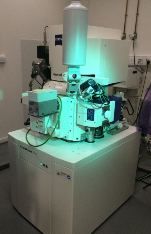
This is an electron microscope
What can you look at using an electron microscope?
Electron microscopes can be used to study a huge range of biological specimens, from tiny molecules such as proteins, to larger structures such as cells or tissues, and everything in between. Electron microscopy can be used to advance our understanding of many different processes, such as how healthy cells grow and develop, how bacteria and viruses infect our cells, how our immune systems fight infectious diseases and cancer, how our brains work and how we age.
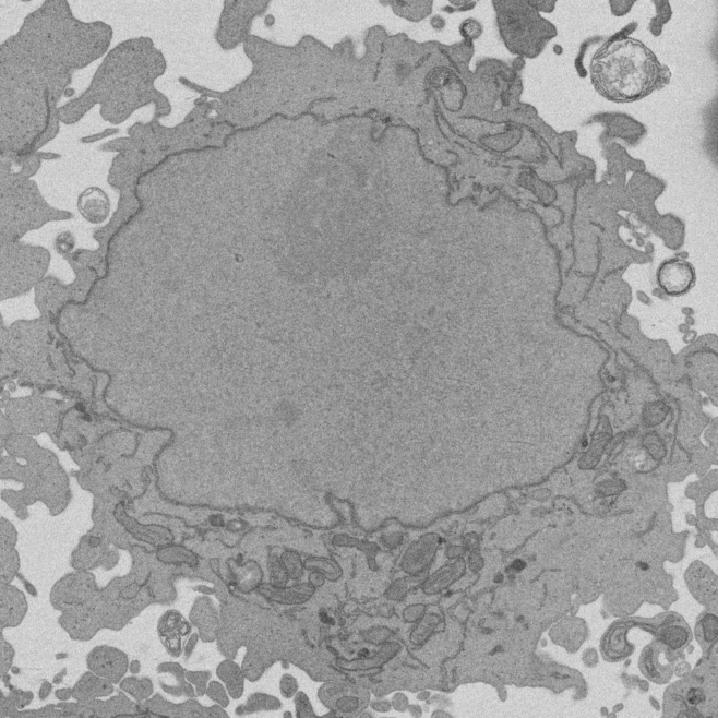
Image of a cell taken with an electron microscope. This particular cell is a HeLa cell, a type of cancer cell that is widely used in scientific research.
How do we study the feature of interest in the image?
An image taken with an electron microscope contains a huge amount of information. The information we want to extract from an image will depend on our particular research question. Until recently though, most electron microscopes could only give a 2D view from a very thin slice through a cell or tissue. This was a problem because humans aren’t very good at visualising 3D structures from a 2D image. Think of the difference between seeing a city on a 2D map, and actually visiting that city and moving through it and seeing how all the roads and buildings and rivers and people interact with each other in 3D.
How can we study specimens in 3D?
We can look at a cell in 3D using different types of microscope, the two we have used here are called a serial block face scanning electron microscope (SBF SEM) and a focused ion beam scanning electron microscope (FIB SEM). In these microscopes, the cell surface is imaged, and then a thin ‘section’ is cut away, and then the cell is imaged again. A diamond knife is used to cut the sections away in the SBF SEM and a highly focused beam of heavy ions is used in the FIB SEM. These microscopes can cut sections so thin that it would take 2,000-20,000 sections to cut through the width of a single human hair! This process of cutting and imaging creates a series of 2D images.
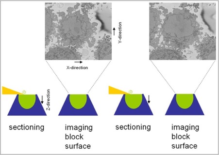
Serial block face scanning electron microscopy
How can we generate a 3D model from lots of 2D images?
One way to convert these 2D images into a 3D model is to draw around the feature(s) of interest. This is known as ‘segmentation’. We also call it ‘advanced colouring in’!

Here's Anne segmenting an image in our lab
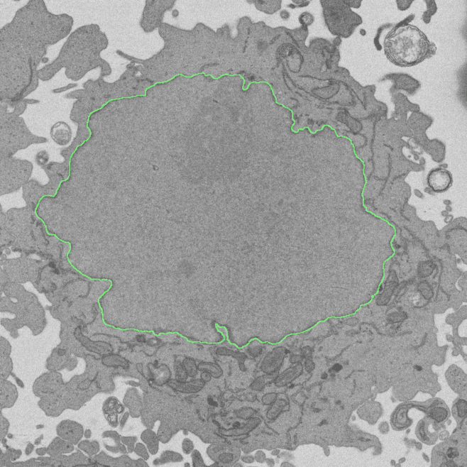
In this image the cell's nucleus is "segmented", with the annotation overlay toggling on and off for clarity
How can this be used to generate a 3D structure?
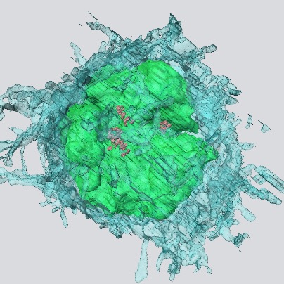
A 3D model of the cell can be created by combining the segmentations from the series of 2D images produced by the serial block face scanning electron microscope. The individual segmentations are stacked together to create a 3D surface. It is much easier for humans to understand this 3D nuclear envelope, and to compare it to diseased nuclear envelopes, than it is to understand the 2D images showing a very small part of the structure. In the example below, you can just make out the individual segmentations as the approximately horizontal ridges on the 3D surface.
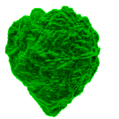
3D reconstruction of 2D segmented images of a nucleus
Why do we need your help?
The process of manual segmentation can take a really long time - it is the major bottleneck in our research - this is why we need your help! While it may look as though computers would be able to perform this process very well already, in practice they are still quite error-prone and the time it takes to perfect the algorithm can be longer than the time to do the segmentations by hand. Small variations in the images, either due to real biological differences or due to slightly different imaging conditions, often render a finely-tuned algorithm ineffective for other data, sometimes meaning that the algorithms need to be optimised for each and every sample!
We hope that this project will advance our ability to analyse biological features using electron microscopy and segmentation. With enough volunteer help we may be able to train computers to segment automatically; this has huge potential to help us understand biology and study disease!
What are our research goals?
Your contribution to this project will help us to understand the structure of the nuclear envelope. Working on our own, we could segment the nuclear envelope of just one cell per week. We need segmentations from hundreds or thousands of cells to understand how the nuclear envelope works in healthy cells, and in cells that have been virus infected, or from tumours, or from patients with rare inherited diseases. That means segmenting tens or hundreds of thousands of individual images.
With your help, we will be able to make large numbers of 3D models of nuclear envelopes for further analysis. Even better, we will be able to use your segmentations to train computers to perform these segmentations automatically. By understanding the 3D structure of the nuclear envelope, we will be able to measure the effect of different diseases on this vitally important organelle, which in turn allows us to work out how new treatments might be created.
If you would like to read more about the nuclear envelope in health and disease, you could have a look at this review paper:
‘Breaching the nuclear envelope in development and disease’ by Hatch & Hetzer (2014).
This project is part of the Etch A Cell Organisation

'Etch A Cell' is one of multiple projects produced by the Etch A Cell team and their collaborators to explore different aspects of cell biology. If you'd like to get involved in some of our other projects, you can find the other Etch A Cell projects on our organisation page here.
The science team behind Etch A Cell are based at the Francis Crick Institute in London, UK. There they work with many other researchers to study different aspects of biology using cutting-edge Electron Microscopes. These microscopes have a very high magnification and resolution, and so can be used to take highly-detailed images of many things, including cells, molecules and tissues. These images can be used to provide us with a richer understanding of biology, which can help us understand the biological changes associated with health and disease.
Recent developments in electron microscopy mean it is possible to collect images automatically, so our team is now producing a huge amount data; looking at a single cell alone can produce several terabytes worth! Although these technological advances mean we can perform our research much faster, this flood of data has caused a data analysis bottleneck, which is why we need your help!
With thanks to support from...


