


Finished! Looks like this project is out of data at the moment!
Thanks to the amazing dedication of our volunteer community, this project is now finished!
You can learn more about Etch A Cell and discover our new projects here
Research
Overview of our research – who are we, what are we studying, and why?
The science team behind Etch A Cell are based at the Francis Crick Institute in London, UK. There they work with many other researchers to study different aspects of biology using cutting-edge Electron Microscopes. These microscopes have a very high magnification and resolution, and so can be used to take highly-detailed images of many things, including cells, molecules and tissues. These images can be used to provide us with a richer understanding of biology, which can help us understand the biological changes associated with health and disease.
Recent developments in electron microscopy mean it is possible to collect images automatically, so our team is now producing a huge amount data; looking at a single cell alone can produce several terabytes worth! Although these technological advances mean we can perform our research much faster, this flood of data has caused a data analysis bottleneck, which is why we need your help!

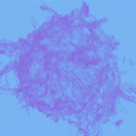
In this project you'll get to see some very high resolution images of cells like this one!
How you can help – colour in cells for science!
To make sense of the images taken by the electron microscope we must carefully analyse them. To extract meaning from the images we segment the features we’re interested in, which means to draw around the bits of the cell that we want to study. We need your help with this task – to colour in different parts of the cell. In this second Etch A Cell project, the cellular feature we would like you to help us segment are the mitochondria.


In this project we need you to spot and draw around mitochondria like these ones (for more examples, see our mitochondria gallery on the Field Guide tab on the right hand side of your screen)
Mitochondria – the powerhouses of your cells
Mitochondria are a type of organelle (a small, specialized structure) found within eukaryotic cells. They are often referred to as the ‘powerhouses’ of the cell as they convert fat, protein and sugar from food into chemical energy. In addition to the production of energy, mitochondria also contribute to other critical biological processes, including the regulation of cell growth and cell death. It is because of their role in a variety of cellular functions that the malfunction of mitochondria is associated with a range of different diseases, which makes them a particularly important organelle to study. Most cells within your body contain mitochondria, but they can vary in number, size, structure and position. In this project, we ask you to simply draw around each mitochondria you see in each image. This effort will help significantly improve our understanding of mitochondrial biology.
Why do we need your help?
The process of manual segmentation can take a really long time - it is the major bottleneck in our research - this is why we need your help! While it may look as though computers would be able to perform this process very well already, in practice they are still quite error-prone and the time it takes to perfect the algorithm can be longer than the time to do the segmentations by hand. Small variations in the images, either due to real biological differences or due to slightly different imaging conditions, often render a finely-tuned algorithm ineffective for other data, sometimes meaning that the algorithms need to be optimised for each and every sample!
We hope that this project will advance our ability to analyse biological features using electron microscopy and segmentation. With enough volunteer help we may be able to train computers to segment automatically; this has huge potential to help us understand biology and study disease!
Further information
For more information about the scientific methods we use in our research, take a look at the FAQ.
This project is part of the Etch A Cell Organisation
'Etch A Cell - Powerhouse Hunt' is one of multiple projects produced by the Etch A Cell team and their collaborators to explore different aspects of cell biology. If you'd like to get involved in some of our other projects, you can find the other Etch A Cell projects on our organisation page here.
The science team behind Etch A Cell are based at the Francis Crick Institute in London, UK. There they work with many other researchers to study different aspects of biology using cutting-edge Electron Microscopes. These microscopes have a very high magnification and resolution, and so can be used to take highly-detailed images of many things, including cells, molecules and tissues. These images can be used to provide us with a richer understanding of biology, which can help us understand the biological changes associated with health and disease.
Recent developments in electron microscopy mean it is possible to collect images automatically, so our team is now producing a huge amount data; looking at a single cell alone can produce several terabytes worth! Although these technological advances mean we can perform our research much faster, this flood of data has caused a data analysis bottleneck, which is why we need your help!
With thanks to support from...


